

Oculoplasty and Periocular Surgery
Oculoplasty is a specialised branch of ophthalmology focused on the eyelids, tear ducts, eye socket, surrounding facial tissues and structures around the eyes. It combines eye care with reconstructive and aesthetic surgery to improve both function and appearance. Oculoplastic procedures may be used to treat drooping eyelids, eyelid malpositions, tear duct blockage orbital disorders, trauma, tumours and socket related problems. Periocular surgery also includes procedures designed to restore symmetry and natural contours around the eyes.
At Viaan Eye & Retina Centre, evaluation begins with understanding the underlying condition so treatment can be planned around vision, comfort, eye protection and appearance.
When Should You Consult an Oculoplasty Specialist?
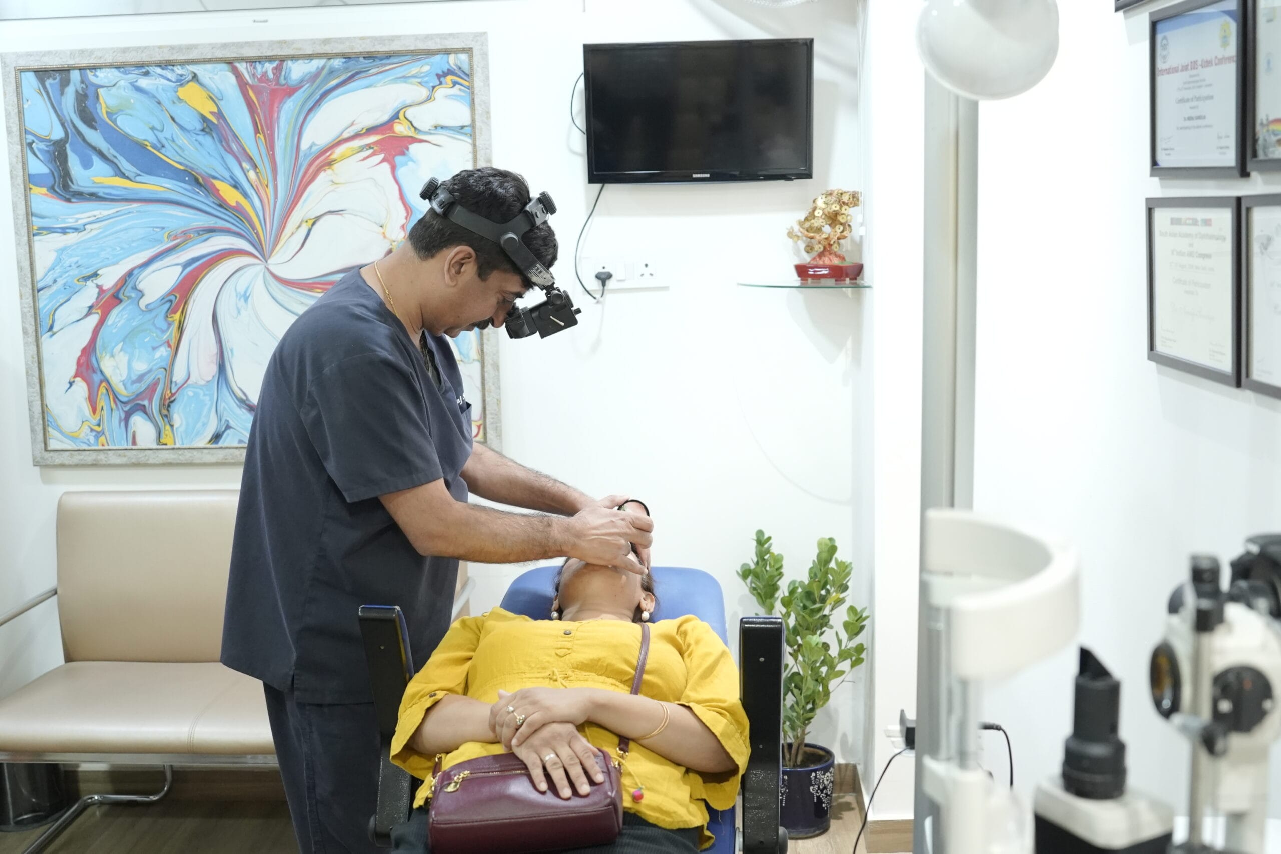
You should consult an oculoplasty specialist when problems involving the eyelids, tear ducts, eye socket or surrounding tissues begin affecting vision, comfort, appearance or normal eye function.
Conditions Affecting the Eyelids
The eyelids do much more than frame the eyes. They help protect the eye surface, spread tears evenly, keep the cornea moist and close properly during sleep. When the eyelids change in position, movement or structure they can affect both comfort and vision. Oculoplasty treatment focuses on correcting these problems while protecting normal eye function and maintaining a natural appearance.
Ptosis or Drooping Eyelid
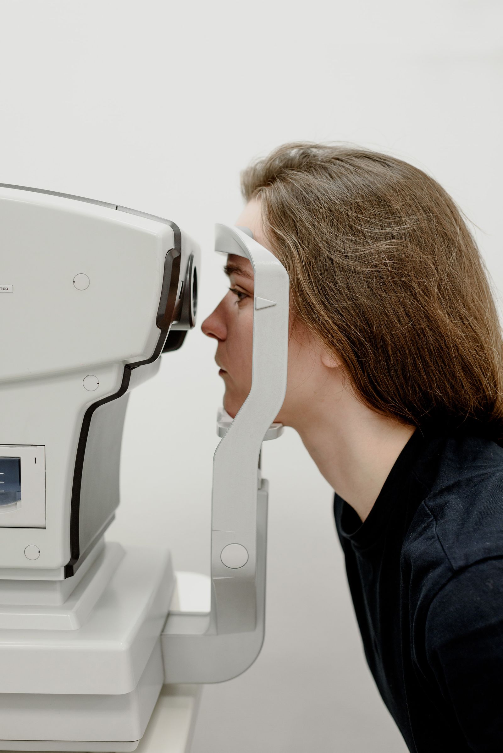
Ptosis occurs when the upper eyelid sits lower than normal. It may be present from birth or develop later because of ageing, muscle weakness, nerve problems, injury or previous eye surgery.
In mild cases, ptosis may mainly affect appearance. In more advanced cases the eyelid can cover part of the pupil and interfere with vision. Children with significant ptosis may also be at risk of visual development problems if the eyelid blocks the visual axis.
Treatment depends on the cause, severity, eyelid muscle strength and amount of visual obstruction.
Entropion
Entropion occurs when the eyelid turns inward, causing the eyelashes and skin to rub against the eye surface.
This can lead to:
- Irritation
- Watering
- Redness
- Foreign body sensation
- Corneal scratches
- Recurrent infection
- Corneal damage in severe cases
It commonly affects the lower eyelid and may develop with ageing, scarring or previous injury. Surgical correction may be recommended when the condition is persistent or damaging the eye surface.
Ectropion
Ectropion is the opposite problem, where the eyelid turns outward and no longer rests properly against the eye.
This can prevent tears from spreading normally across the eye surface and may cause:
- Excessive watering
- Dryness
- Irritation
- Redness
- Exposure of the inner eyelid
- Recurrent eye surface problems
Ectropion may occur because of ageing, facial nerve weakness, scarring, trauma or previous surgery.
Eyelid Retraction
Eyelid retraction occurs when the upper or lower eyelid is pulled farther away from its normal position, exposing more of the eye.
Patients may experience dryness, irritation, watering, sensitivity to light or difficulty closing the eye completely. It can be associated with thyroid eye disease, previous surgery, trauma or scarring.
Treatment is planned according to the underlying cause and degree of exposure.
Lagophthalmos
Lagophthalmos means the eyelids cannot close completely.
When the eye remains partly open, especially during sleep the cornea may become dry and exposed. Patients may experience:
- Burning
- Gritty sensation
- Watering
- Redness
- Blurred vision
- Corneal irritation
Possible causes include facial nerve weakness, trauma, eyelid surgery, scarring or orbital disease.
Eyelid Lumps and Cysts
Different types of lumps can develop on or within the eyelids. Some are harmless, while others require closer evaluation.
Common examples include:
- Chalazion
- Stye
- Sebaceous cyst
- Papilloma
- Skin tags
- Other benign eyelid lesions
A lump that does not resolve, keeps returning, changes in appearance, causes bleeding or affects the eyelid margin should be examined properly.
Eyelid Tumours
Tumours can develop in the eyelid skin, glands or surrounding tissues. Some are benign, while others can be malignant.
Warning signs may include:
- A persistent or enlarging lump
- Loss of eyelashes
- Ulceration
- Bleeding
- Irregular eyelid margin
- Change in skin colour
- Recurrent lesion at the same site
Suspected tumours may require biopsy, surgical removal and reconstruction depending on their size and location.
Blepharochalasis and Excess Eyelid Skin
With age the skin and tissues around the upper and lower eyelids may become loose or heavy.
Excess upper eyelid skin can sometimes extend over the eyelid crease and interfere with the upper field of vision. Lower eyelid changes may create puffiness or bags.
When excess tissue affects function or appearance, blepharoplasty may be considered.
Eyelid Scarring
Scarring after trauma, burns, infection or previous surgery can alter eyelid position and movement.
Scar tissue may cause:
- Eyelid shortening
- Retraction
- Entropion
- Ectropion
- Poor eyelid closure
- Facial asymmetry
Reconstructive oculoplasty may be needed to restore eyelid position and protect the eye.
Eyelid Problems After Facial Nerve Palsy
Weakness of the facial nerve can reduce the ability to blink or close the eye properly.
This may leave the cornea exposed and cause dryness, watering, irritation or even corneal injury. Management can involve lubrication, temporary protective measures, eyelid weights or reconstructive procedures depending on severity.
How We Evaluate Eyelid and Orbital Problems
Eyelid and orbital conditions can affect much more than appearance. They may interfere with eye closure, tear drainage, corneal protection, eye movement, vision and the position of the eye within the socket. A detailed oculoplasty evaluation therefore looks at both function and facial symmetry before deciding whether observation, medical treatment, imaging or surgery is required.
Medical and Surgical History
The evaluation begins by understanding the patient’s symptoms, when they started and whether they are getting worse. Previous eye surgery, facial trauma, thyroid disease, infections, allergies, tumours and other medical conditions are also reviewed.
Eyelid Position and Function
The upper and lower eyelids are carefully examined for:
- Drooping or ptosis
- Eyelid retraction
- Entropion or inward turning
- Ectropion or outward turning
- Incomplete eye closure
- Eyelid symmetry
- Eyelid muscle strength
- Blink function
These measurements help determine whether the eyelid is protecting the eye properly.
Eye Surface and Corneal Assessment
Poor eyelid position can expose the cornea and disturb the tear film. The eye surface is therefore checked for dryness, irritation, staining, inflammation or exposure related damage.
Eye Position and Orbital Examination
The specialist assesses whether the eye is positioned normally within the socket. Signs such as bulging, displacement, sinking, swelling or asymmetry can point toward an orbital condition.
Measurements may also be taken to compare the position of both eyes.
Eye Movement and Double Vision Assessment
Patients with orbital disease may experience restricted eye movement or double vision. Eye movements are checked in different directions to determine whether muscles, nerves or tissues inside the orbit are being affected.
Tear Drainage Evaluation
If the main concern is excessive watering, recurrent discharge or tear duct blockage the tear drainage pathway may be examined. Additional tests may be performed to identify the location and severity of the obstruction.
Examination of Eyelid Lumps or Lesions
Any eyelid lump, cyst, ulcer, pigmentation or abnormal growth is assessed for:
- Size
- Location
- Surface appearance
- Eyelash loss
- Bleeding or ulceration
- Rate of growth
- Involvement of surrounding tissues
Suspicious lesions may require further investigation or biopsy.
Orbital Imaging
When deeper structures cannot be evaluated adequately during examination, imaging may be advised. CT or MRI scans can help assess the eye socket, muscles, optic nerve, surrounding tissues, fractures, inflammation or suspected masses.
Clinical Photography and Measurements
Photographs and precise eyelid measurements may be taken before treatment. These provide a baseline for surgical planning and allow changes to be compared during follow up.
Imaging and Diagnostic Tests Used in Oculoplasty
Oculoplasty involves structures around the eye, including the eyelids, tear drainage system orbit, eye muscles, surrounding soft tissues and facial structures. Because many of these areas cannot be fully assessed by looking at the surface alone, specialised measurements, imaging and diagnostic tests may be required. The tests chosen depend on the patient’s symptoms and the suspected condition.
CT Scan of the Orbit
A CT scan provides detailed images of the bony orbit and surrounding structures. It is particularly useful when evaluating:
- Orbital fractures after trauma
- Bone abnormalities
- Foreign bodies
- Sinus related orbital disease
- Certain orbital masses
- Thyroid related orbital changes
CT imaging can help determine the location, size and extent of a problem before treatment or surgery is planned.
MRI of the Orbit
MRI provides high detail images of the soft tissues inside and around the eye socket. It can be especially useful for assessing:
- Orbital tumours
- Optic nerve abnormalities
- Eye muscle disorders
- Inflammation within the orbit
- Vascular lesions
- Deeper soft tissue changes
MRI does not use ionising radiation and may provide better soft tissue detail than CT in selected conditions.
Orbital Ultrasonography
Ultrasound may be used to evaluate structures behind or around the eye when additional information is needed. It can help assess certain orbital masses, cysts, inflammation and structural abnormalities.
Exophthalmometry
Exophthalmometry measures how far the eye projects forward from the orbit.
It is particularly useful when assessing proptosis or bulging eyes, including cases associated with thyroid eye disease or orbital masses. Measurements from both eyes can also be compared during follow up.
Eyelid Measurements
Precise measurements help evaluate eyelid position and function. These may include:
- Upper eyelid height
- Margin reflex distance
- Levator muscle function
- Eyelid crease position
- Degree of ptosis
- Eyelid symmetry
- Amount of incomplete closure
These measurements are especially important when planning ptosis correction or reconstructive eyelid surgery.
Tear Drainage Tests
Patients with constant watering or recurrent tear sac infections may need evaluation of the lacrimal drainage system.
Tests may include:
- Tear drainage assessment
- Syringing and probing
- Fluorescein dye testing
- Assessment of punctal openings
- Imaging of the tear drainage pathway in selected cases
These tests help determine whether the blockage is partial or complete and where it is located.
Visual Acuity and Eye Examination
Vision is assessed because eyelid or orbital conditions can sometimes interfere with sight. A complete eye examination may also include evaluation of the cornea, pupils, optic nerve, eye pressure and ocular surface.
Eye Movement and Double Vision Testing
Patients with orbital disease may develop restricted eye movement or diplopia. Eye movements are checked in different directions to identify possible involvement of the extraocular muscles, nerves or orbital tissues.
Visual Field Testing
When drooping eyelids or excess upper eyelid skin obstruct peripheral vision, a visual field test can measure how much the eyelid is affecting the patient’s field of sight.
Clinical Photography
Standardised photographs may be taken before treatment to document:
- Eyelid position
- Facial symmetry
- Swelling
- Lesions
- Proptosis
- Surgical planning measurements
They also allow objective comparison during follow up.
Biopsy and Histopathology
If an eyelid or orbital lesion appears suspicious, a small tissue sample may be removed and examined under a microscope. Biopsy helps determine whether the lesion is benign, inflammatory or malignant and guides further treatment.
How We Choose the Right Oculoplasty Treatment
Oculoplasty treatment is never based on appearance alone. The right approach depends on which structure is affected, how much function is disturbed the underlying cause and whether the problem is medical, reconstructive or aesthetic.
Treatment begins by understanding whether the problem involves the eyelid, tear duct orbit, eye socket, surrounding tissues or a combination of these areas. Conditions such as ptosis, entropion, ectropion, blocked tear ducts orbital disease, trauma and eyelid lesions all require different treatment approaches.
The specialist first looks at whether the condition is affecting:
- Eye closure
- Corneal protection
- Tear drainage
- Vision
- Eye movement
- Comfort
- Peripheral visual field
- Position of the eye within the socket
A treatment plan is then created to restore normal function while maintaining a balanced and natural appearance.
Not every oculoplasty condition requires surgery. Mild problems may be managed with observation, medicines, lubrication or regular monitoring.
Surgery may be considered when the condition begins to interfere with eye protection, vision, tear drainage, comfort or normal eyelid position.
For conditions such as ptosis, detailed measurements are used to understand how low the eyelid sits and how well the lifting muscle works.
This helps determine which surgical technique may provide the most appropriate correction.
If the eyelids do not close properly the cornea may become dry or exposed. In these cases, treatment planning must first consider how well the eye surface is being protected.
The priority is to prevent ongoing irritation or corneal damage.
For persistent watering or recurrent tear sac infections the tear drainage pathway is examined to locate the blockage.
Depending on the cause and location, treatment may involve a minor procedure, tear duct surgery or DCR surgery to create a new drainage pathway.
Conditions involving the eye socket often require imaging such as CT or MRI.
These scans help identify:
- Orbital inflammation
- Thyroid eye disease
- Tumours
- Fractures
- Muscle involvement
- Deep tissue abnormalities
Treatment may then involve medicines, monitoring orbital surgery or reconstructive procedures.
Eyelid lumps are assessed according to their size, location, growth pattern, appearance and effect on surrounding tissues.
Simple benign lesions may only require observation or removal, while suspicious growths may need biopsy, complete excision and eyelid reconstruction.
Previous eye surgery, burns, facial injury or scarring can change normal eyelid and orbital anatomy.
These cases often require a more customised reconstructive approach because treatment may need to restore both tissue position and function.
Depending on the condition, treatment may include:
- Lubricating eye drops
- Anti inflammatory medicines
- Antibiotics
- Management of thyroid or systemic disease
- Observation and follow up
- Eyelid correction
- Ptosis surgery
- DCR surgery
- Orbital surgery
- Tumour removal
- Socket reconstruction
- Reconstructive or aesthetic procedures
For periocular aesthetic procedures, facial symmetry, eyelid position, skin quality and the patient's expectations are considered together.
The goal is not simply to change appearance but to preserve normal eyelid movement, eye protection and a natural result.
Choose the Right Oculoplasty Treatment for Your Eyes
What Happens During Oculoplasty Surgery
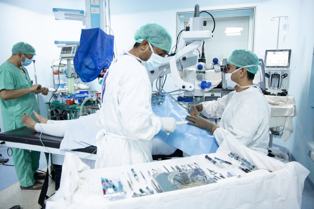
Oculoplasty surgery is planned according to the structure being treated, whether it involves the eyelid, tear duct orbit, eye socket or surrounding tissues. The exact procedure varies from patient to patient but the aim is to restore normal function while maintaining natural eye protection, comfort, symmetry and appearance.
1. Pre Surgical Assessment
Before surgery the oculoplasty specialist reviews the diagnosis, eyelid position, eye movements, tear drainage, socket condition and overall eye health. Photographs, measurements and imaging such as CT or MRI may be used when deeper orbital structures are involved.
The surgical plan is then tailored to the condition being treated.
2. Anaesthesia
Oculoplasty procedures may be performed under local anaesthesia, local anaesthesia with sedation or general anaesthesia, depending on the complexity of the surgery, patient age and area being treated.
Anaesthesia helps keep the patient comfortable throughout the procedure.
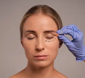
3. Precise Surgical Marking
For eyelid procedures, careful markings may be made before surgery to guide incision placement and help maintain proper symmetry.
The surgeon considers:
- Natural eyelid folds
- Eyelid height
- Position of the eyelashes
- Eye closure
- Facial symmetry
- Amount of tissue requiring correction
4. Treatment of the Underlying Problem
The surgical technique depends on the diagnosis.
- Ptosis: The eyelid lifting muscle may be tightened, shortened or repositioned to raise the eyelid and improve the visual field.
- Entropion or Ectropion: The eyelid tissues are repositioned and tightened so the lid rests correctly against the eye surface.
- Tear Duct Blockage: Procedures such as DCR may be performed to create a new pathway for tears to drain from the eye into the nose.
- Eyelid Lesions or Tumours: The abnormal tissue may be removed and the eyelid reconstructed when necessary to restore function and appearance.
- Orbital Conditions: Surgery may involve treatment of orbital masses, fractures, inflammation related structural problems or tissue compression depending on the underlying disease.
- Socket Reconstruction: Scarred or contracted tissue may be repaired or reconstructed to improve socket shape and support future prosthetic fitting.
5. Tissue Reconstruction and Alignment
Once the main problem is corrected the surgeon carefully restores tissue position and alignment.
Special attention is given to maintaining:
- Proper eyelid closure
- Corneal protection
- Natural contour
- Symmetry between both eyes
- Normal tear flow
- Stable eyelid position
In reconstructive procedures, surrounding tissue may be repositioned or grafted when required.
6. Fine Suturing and Wound Closure
Small sutures are used where necessary to close the surgical area. Incisions are often planned along natural eyelid folds or existing skin lines to make healing as discreet as possible.
Some stitches may dissolve naturally, while others may need removal during follow up.
7. Immediate Post Surgery Care
After surgery the treated area is checked for swelling, bleeding, eyelid position, eye movement and overall eye safety.
Patients may be prescribed:
- Antibiotic ointment or drops
- Lubricating eye drops
- Anti inflammatory medication
- Cold compresses
- Pain relief medication when required
8. Recovery and Follow Up
Mild swelling, bruising or temporary discomfort can occur during the early recovery period. Follow up visits allow the surgeon to monitor healing, remove stitches if needed and check eyelid function, symmetry and eye surface health.
Recovery and Aftercare Following Oculoplasty
Recovery after oculoplasty depends on the type of procedure performed, whether it involves the eyelids, tear ducts orbit, eye socket or reconstructive tissues. Some patients recover within a few days, while more complex orbital or reconstructive procedures may require several weeks. Proper aftercare helps reduce swelling, protect the eye, support healing and improve the final functional and cosmetic outcome.
Swelling and Bruising
Mild to moderate swelling and bruising around the eyes are common after many oculoplasty procedures. These usually improve gradually over the first one to two weeks.
Cold compresses may be advised during the early recovery period to help reduce swelling and discomfort.
Eye Drops, Ointments and Medicines
Depending on the surgery, your doctor may prescribe:
- Antibiotic eye drops or ointment
- Lubricating eye drops
- Anti inflammatory medication
- Pain relief medicines
- Oral antibiotics when required
These should be used exactly as advised and should not be stopped without medical guidance.
Keeping the Surgical Area Clean
Patients are usually advised to keep the surgical area clean and avoid rubbing or pressing around the operated eye.
Any crusting or discharge should be cleaned only in the manner recommended by the clinical team.
Protecting the Eye During Healing
The eye and surrounding tissues may be more sensitive during recovery. Patients may be advised to:
- Wear sunglasses outdoors
- Avoid dust and smoke
- Avoid eye makeup temporarily
- Protect the operated area from accidental rubbing
- Avoid direct pressure on the eyelids or socket
Activity Restrictions
Strenuous activity can increase swelling or bleeding during the early healing period.
Patients may be advised to temporarily avoid:
- Heavy lifting
- Vigorous exercise
- Bending for long periods
- Swimming
- Contact sports
- Activities that put pressure around the eyes
The timing for returning to normal activity depends on the procedure performed.
Sleeping Position
Sleeping with the head slightly elevated may help reduce swelling during the first few days. Your surgeon may also advise avoiding pressure on the operated side.
Stitches and Wound Care
Some oculoplasty procedures use dissolvable stitches, while others require removal during a follow up visit.
Patients should avoid pulling, scratching or applying products directly over the incision until advised that healing is adequate.
Follow Up Appointments
Follow up visits are important for monitoring:
- Wound healing
- Eyelid position
- Eye closure
- Corneal protection
- Tear drainage
- Swelling and inflammation
- Symmetry
- Vision and eye movement
Adjustments to medicines or additional treatment may be made depending on recovery.
When Can You Return to Work?
Return to work depends on the procedure and type of job. Patients undergoing minor eyelid procedures may return sooner, while orbital or reconstructive surgery may require a longer recovery period.
Bruising and swelling may remain visible even when the patient feels comfortable enough to resume routine activities.
Watch for Warning Signs
Patients should contact their eye specialist promptly if they develop:
Why Choose Viaan Eye and Retina Centre?
- Specialised expertise in eyelid, tear duct and orbital conditions
- Personalised oculoplasty treatment after a detailed eye examination
- Care focused on eye function, comfort and natural appearance
- Precise treatment planning for delicate areas around the eyes
- Clear guidance about the procedure, recovery and expected results
- Regular follow ups to support comfortable healing and lasting improvement

Could Oculoplasty Treatment Help You?
You may benefit from oculoplasty treatment if an eyelid, tear duct or eye socket concern is affecting your comfort, vision, appearance or confidence.
Expert Led Care from Evaluation to Final Restoration
Every case is different, so we take time to understand the concern properly and plan care that feels clear, thoughtful and reassuring.
1. First Consultation: We listen to your concerns, review your medical history and understand how the eyelid, tear duct or eye socket problem affects your comfort or appearance.
2. Detailed Examination: The doctor checks your eyelids, tear drainage, eye movement, vision and surrounding facial structures to identify the exact cause of the problem.
3. Treatment Planned for You: Your oculoplasty treatment is selected according to the diagnosis your daily needs, overall health and the results you hope to achieve.
4. Procedure and Aftercare: The procedure is performed with close attention to the delicate eye area. You receive simple guidance on medicines, precautions and recovery.
5. Follow Up Visits: Regular check ups help the doctor review healing, eyelid position, comfort and progress while addressing any concerns during recovery.
Possible Risks and Safety Considerations
Oculoplasty procedures are generally planned carefully to protect both eye function and appearance but like any surgery they can involve risks. The type and likelihood of complications depend on the procedure performed the patient’s eye health, medical history, healing response and the complexity of the condition being treated.
Swelling and Bruising
Temporary swelling and bruising are common after eyelid and periocular surgery. These usually settle gradually as healing progresses.
Bleeding or Haematoma
A small amount of bleeding may occur after surgery. In rare cases, blood can collect within the tissues and may require urgent medical attention, particularly if it causes severe pain, swelling or vision changes.
Infection
Although uncommon, infection can develop around the surgical site. Increasing redness, warmth, swelling, discharge, fever or worsening pain should be reported promptly.
Temporary Blurred Vision
Some patients may notice temporary blurred vision because of swelling, ointments, tear film disturbance or dryness during the early recovery period.
Dryness and Eye Surface Irritation
Changes in blinking or eyelid position can temporarily affect tear distribution. Lubricating drops may be recommended to keep the eye surface comfortable during recovery.
Difficulty Closing the Eye
After certain eyelid procedures, complete eye closure may temporarily become difficult because of swelling or tissue adjustment. Persistent exposure requires review to protect the cornea.
Eyelid Asymmetry
Minor differences between the two eyelids may remain during healing. In some cases, noticeable asymmetry may persist and require further assessment or correction.
Under Correction or Over Correction
Procedures for ptosis or eyelid position may occasionally result in the eyelid sitting slightly higher or lower than intended. Revision treatment may sometimes be considered after healing is complete.
Scarring
Oculoplasty incisions are usually positioned carefully around natural eyelid folds where possible but every surgical procedure can leave some degree of scarring. Scar appearance generally changes with time.
Changes in Eyelid Position
Rarely, healing or scar formation can lead to problems such as ectropion, entropion, eyelid retraction or altered eyelid contour, which may require additional treatment.
Double Vision or Eye Movement Changes
Orbital surgery can occasionally affect the muscles controlling eye movement, resulting in temporary or persistent double vision. The risk depends greatly on the condition and type of surgery performed.
Reduced Sensation
Temporary numbness or altered sensation around the eyelids, forehead or cheek may occur when small sensory nerves are affected during surgery. This often improves gradually.
Tear Drainage Problems
Following tear duct procedures, blockage can occasionally recur or watering may continue despite treatment. Further examination may be required if symptoms persist.
Risks Related to Orbital Surgery
Procedures involving deeper structures of the eye socket carry additional risks because the optic nerve, eye muscles, blood vessels and other delicate tissues are located within a confined space.
Complications are uncommon but can be more serious and require careful surgical planning and monitoring.
Anaesthesia Related Risks
Local, sedation or general anaesthesia may carry risks depending on the patient’s medical condition. Health history, current medicines, allergies and previous anaesthesia experiences should therefore be discussed before surgery.
Protecting Safety Before Surgery
A safe treatment plan begins with proper evaluation. Before recommending surgery the specialist may review:
- Eye and eyelid function
- Corneal and ocular surface health
- Vision and eye movements
- Tear drainage
- Medical conditions and medications
- Previous eye or facial surgery
- Imaging findings when required
- Expected functional and cosmetic outcome
Let Your Eyes Feel and Look More Balanced
Get a clear diagnosis and personalised oculoplasty treatment for eyelid, tear duct or eye socket concerns affecting your comfort or appearance.
- Specialised Oculoplasty Care
- Functional & Aesthetic Precision
- Personalised Treatment Planning
- Careful Recovery Support
Frequently Asked Questions
1. What is oculoplasty treatment?
Oculoplasty treatment manages problems affecting the eyelids, tear ducts, eye socket and nearby tissues, improving eye protection, comfort, function, appearance or facial balance when needed.
2. Which conditions can oculoplasty treat?
Treatment may help with drooping eyelids, inward or outward turning lids, blocked tear ducts, eyelid growths, injuries, socket concerns and congenital eyelid defects in children.
3. Is oculoplasty only a cosmetic procedure?
Oculoplasty may be medical, reconstructive, cosmetic or a combination. The purpose depends on whether the concern affects vision, comfort, protection, appearance or confidence over time.
4. How long does recovery take after oculoplasty?
Recovery varies with the procedure. Mild swelling or bruising is common, while many patients gradually resume routine activities by following their surgeon’s aftercare advice carefully.
5. How should I choose an oculoplasty specialist?
Choose a specialist experienced in eyelid, tear duct and orbital conditions who provides careful evaluation, clear guidance, personalised planning and dependable follow up after treatment ends.