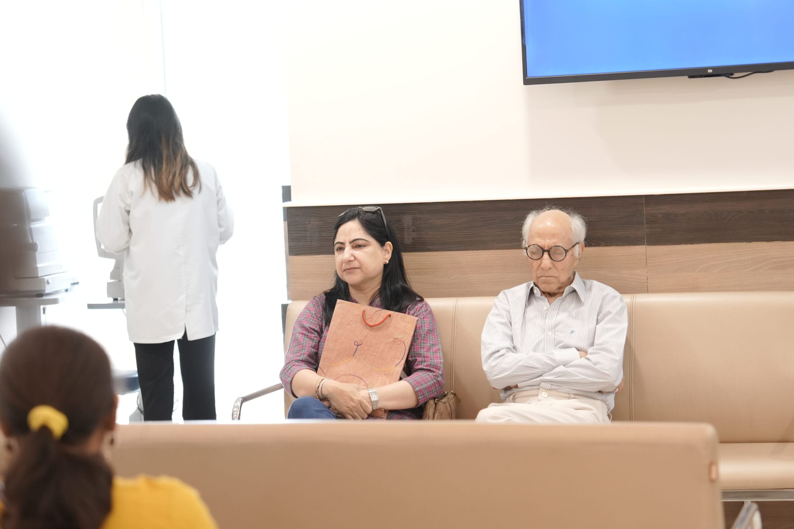


Why Glaucoma Is Often Called the Silent Thief of Sight
Glaucoma is often called the “silent thief of sight” because it can damage vision slowly and without obvious warning signs. In many cases, especially primary open angle glaucoma, the disease begins by affecting peripheral or side vision. Because central vision may remain clear for a long time, a person may not realise that any vision has been lost.
The damage happens at the optic nerve, which carries visual information from the eye to the brain. Once nerve fibres are permanently damaged, the vision already lost usually cannot be restored. This is why detecting glaucoma before noticeable symptoms appear is so important.
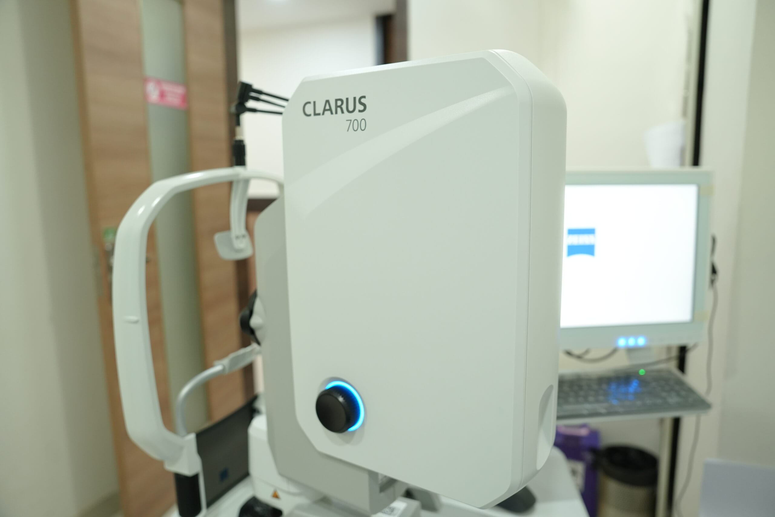
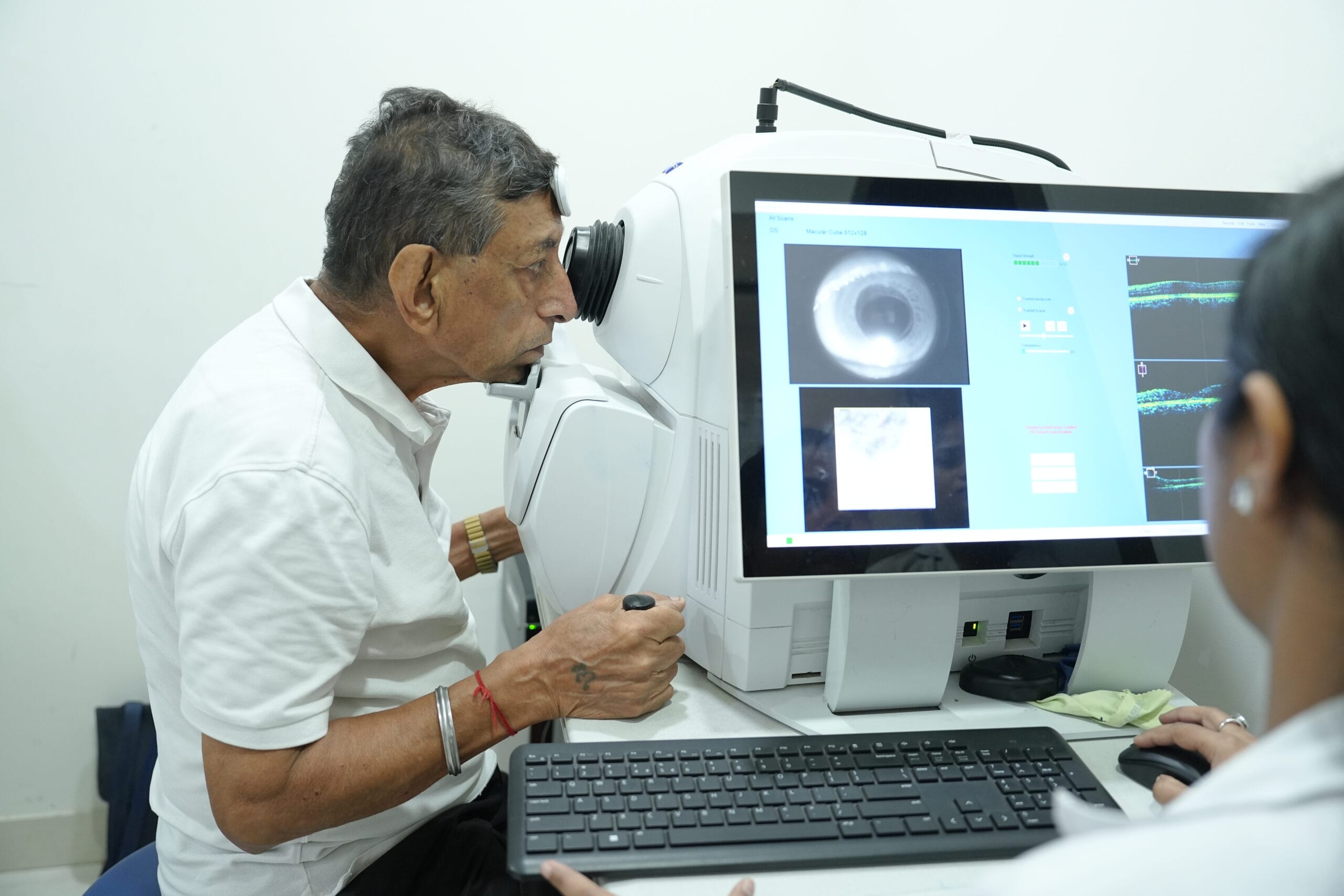
Eye pressure is one of the major factors associated with glaucoma but a normal pressure reading does not always rule it out. A complete glaucoma assessment may include intraocular pressure measurement, optic nerve examination, OCT imaging, visual field testing, corneal thickness measurement and gonioscopy.
As glaucoma progresses, peripheral vision may gradually narrow and, in advanced stages, can lead to severe visual impairment.
At Viaan Eye & Retina Centre, regular glaucoma screening helps identify early optic nerve changes, monitor progression and begin treatment before significant vision loss develops.
Different Types of Glaucoma
Glaucoma is not a single disease. It is a group of eye conditions that damage the optic nerve, the structure that carries visual information from the eye to the brain. Eye pressure plays an important role in many types of glaucoma but glaucoma can also develop when eye pressure is within the normal range. Understanding the type of glaucoma is important because the pattern of damage, speed of progression and treatment approach can differ considerably.
Primary Open Angle Glaucoma
Primary open angle glaucoma is the most common form of glaucoma. It usually develops slowly and often causes no noticeable symptoms during its early stages.
In this condition, the drainage angle between the iris and cornea remains open but the eye's drainage system becomes less efficient. Fluid gradually builds up, which can increase intraocular pressure and place stress on the optic nerve.
Peripheral vision is usually affected first. Because central vision may remain clear for a long time, many people do not realise they have glaucoma until significant optic nerve damage has already occurred.
Regular eye pressure checks, optic nerve examination, OCT imaging and visual field testing are therefore important for early detection.
Angle Closure Glaucoma
Angle closure glaucoma occurs when the drainage angle of the eye becomes narrow or completely blocked, preventing fluid from leaving the eye normally.
It can develop gradually but in some cases the angle closes suddenly and causes a rapid rise in eye pressure. This is known as an acute angle closure attack and requires urgent treatment.
Symptoms may include:
- Severe eye pain
- Sudden blurred vision
- Headache
- Redness of the eye
- Halos around lights
- Nausea or vomiting
- Sudden reduction in vision
Unlike open angle glaucoma, acute angle closure may produce dramatic symptoms and should be treated as an eye emergency.
Normal Tension Glaucoma
In normal tension glaucoma, characteristic optic nerve damage and visual field loss occur even though the measured eye pressure remains within what is traditionally considered the normal range.
The exact reason is not always clear. Some people may have an optic nerve that is particularly sensitive to pressure, while reduced blood supply to the optic nerve may also contribute.
Because the eye pressure may appear normal, diagnosis relies heavily on careful examination of the optic nerve, retinal nerve fibre layer and visual field.
Treatment usually focuses on lowering eye pressure further to reduce additional stress on the optic nerve.
Ocular Hypertension
Ocular hypertension is not technically glaucoma but it is closely related and deserves monitoring.
In ocular hypertension, eye pressure is higher than normal but there is no detectable optic nerve damage or visual field loss.
Not everyone with raised eye pressure develops glaucoma. The risk depends on several factors, including:
- Level of eye pressure
- Corneal thickness
- Age
- Family history
- Optic nerve appearance
- Other eye and medical conditions
Regular monitoring helps identify whether glaucoma begins to develop over time.
Secondary Glaucoma
Secondary glaucoma develops because another eye condition, injury, medication or disease interferes with the eye's normal fluid drainage.
Treatment usually involves controlling both the glaucoma and the underlying cause.
Several important forms fall within this category.
Neovascular Glaucoma
Neovascular glaucoma develops when abnormal new blood vessels grow over the iris and drainage angle.
These vessels can block the eye's drainage system and cause a significant increase in eye pressure.
It is often associated with conditions that reduce blood supply to the retina, particularly:
- Advanced diabetic retinopathy
- Retinal vein occlusion
- Other severe retinal vascular disorders
Treatment may involve controlling eye pressure while also treating the underlying retinal disease with injections, retinal laser or other procedures.
Uveitic Glaucoma
Uveitis is inflammation inside the eye. In some patients, inflammation can interfere with the drainage system and increase eye pressure.
Glaucoma may develop because of:
- The inflammation itself
- Scarring inside the drainage angle
- Long term use of steroid medicines used to control inflammation
Managing uveitic glaucoma can be complex because doctors need to control both eye inflammation and intraocular pressure.
Steroid Induced Glaucoma
Some people experience an increase in eye pressure after using steroid medication.
This may occur with:
- Steroid eye drops
- Oral steroids
- Injections
- Inhaled steroids
- Skin preparations used around the eyes
The response varies from person to person. Patients requiring long term steroid treatment may therefore need regular eye pressure monitoring.
Pigmentary Glaucoma
Pigmentary glaucoma occurs when pigment granules from the back surface of the iris are released into the eye.
These particles can accumulate within the drainage system and make it harder for fluid to leave the eye, resulting in increased pressure.
It may develop from a condition known as pigment dispersion syndrome. Not everyone with pigment dispersion develops glaucoma but regular monitoring is important when eye pressure begins to rise.
Pseudoexfoliative Glaucoma
Pseudoexfoliation syndrome causes abnormal microscopic material to accumulate on structures inside the eye, including the natural lens and drainage system.
When this material blocks fluid drainage and raises eye pressure enough to damage the optic nerve, it is called pseudoexfoliative glaucoma.
Eye pressure in this condition can sometimes fluctuate considerably, making regular monitoring particularly important.
Traumatic Glaucoma
An eye injury can damage the drainage system and cause glaucoma either immediately or many years later.
It may occur after:
- Blunt eye trauma
- Penetrating injuries
- Internal bleeding
- Damage to the drainage angle
- Previous eye surgery
Because glaucoma may develop long after the original injury, anyone with a history of significant eye trauma should inform their ophthalmologist.
Congenital and Childhood Glaucoma
Congenital glaucoma is a rare form that develops in infants or young children because the drainage system of the eye has not formed normally.
Possible signs include:
- Excessive tearing
- Light sensitivity
- Cloudiness of the cornea
- Enlarged appearance of the eye
- Frequent eye squeezing or blinking
Unlike most adult glaucoma, childhood glaucoma often requires surgical treatment to improve drainage and protect the developing optic nerve.
Chronic Angle Closure Glaucoma
Not all angle closure glaucoma develops suddenly. In chronic angle closure, the drainage angle gradually becomes narrower and closes over time.
Patients may have few or no symptoms while optic nerve damage slowly progresses. Gonioscopy and detailed examination of the drainage angle are therefore important in people with narrow angles.
Mixed Mechanism Glaucoma
Some patients have more than one mechanism contributing to glaucoma. For example, a person may have both narrowing of the drainage angle and reduced function of the remaining open drainage system.
These cases are described as mixed mechanism glaucoma and treatment is planned according to the combination of factors affecting eye pressure and optic nerve health.
Glaucoma Eye Drops and Medical Management
For many people with glaucoma, treatment begins with medicated eye drops designed to lower intraocular pressure (IOP). These medicines do not restore vision that has already been lost but they can help reduce further stress on the optic nerve and slow progression of the disease.
The treatment plan depends on the type of glaucoma, current eye pressure, degree of optic nerve damage, visual field changes, age, general health and how the eye responds to treatment. Some patients may need one medicine, while others require a combination of drops to reach their individual target eye pressure.

How Glaucoma Eye Drops Work
Glaucoma medicines generally lower eye pressure in one of two ways:
- Reducing the amount of fluid produced inside the eye
- Helping fluid drain more efficiently from the eye
Using Eye Drops Correctly
Correct technique can improve how much medication reaches the eye.
Patients are generally advised to:
- Wash their hands before using the drops
- Avoid touching the bottle tip to the eye or eyelashes
- Place the prescribed number of drops into the lower eyelid
- Close the eye gently after application
- Avoid repeatedly blinking or squeezing the eyelids
- Follow the correct interval when using more than one type of eye drop
- Use medication at approximately the same time every day
Monitoring Side Effects
Glaucoma medicines are generally well tolerated but side effects can occur depending on the medication.
Possible effects may include:
- Eye redness
- Burning or stinging
- Dryness
- Blurred vision immediately after application
- Changes in eyelash growth
- Darkening around the eyelids or iris with certain medicines
- Fatigue or dizziness
- Changes in heart rate or breathing with some medication classes
Early Signs and Symptoms You Should Know
Glaucoma can develop without obvious symptoms, especially in its early stages. That is why regular eye examinations are important, particularly for people with higher risk factors. Symptoms can vary depending on the type of glaucoma.
Side vision may slowly reduce over time, often without being noticed until the disease has progressed.
Some people may find it harder to see clearly in dim lighting or move comfortably between bright and dark environments.
Changes in clarity, contrast or overall visual comfort may become noticeable as glaucoma progresses.
Coloured rings or halos around lights can occur, particularly when eye pressure rises significantly.
Sudden or severe eye pain can occur in acute angle closure glaucoma and requires urgent medical attention.
A suddenly red eye accompanied by pain or blurred vision may indicate a rapid increase in eye pressure.
Rapid loss of visual clarity can occur during an acute glaucoma attack and should not be ignored.
Severe headache, especially around one eye, may accompany a sudden rise in intraocular pressure.
Acute angle closure glaucoma can sometimes cause nausea or vomiting along with intense eye pain and blurred vision.
As optic nerve damage progresses, peripheral vision may become severely restricted, leaving only a smaller central field of vision.

What We Measure During a Glaucoma Evaluation
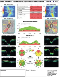
A glaucoma evaluation looks beyond eye pressure, combining structural and functional tests to understand optic nerve health and detect early damage.
Choose the Right Glaucoma Treatment for Your Eyes
Glaucoma Eye Drops
Eye drops are often the first step in eye glaucoma treatment. They help lower eye pressure either by reducing fluid production or improving drainage.
Oral Medicines
In some cases, medicines taken by mouth may be added when eye pressure needs better control. Your glaucoma specialist doctor decides whether they are needed.
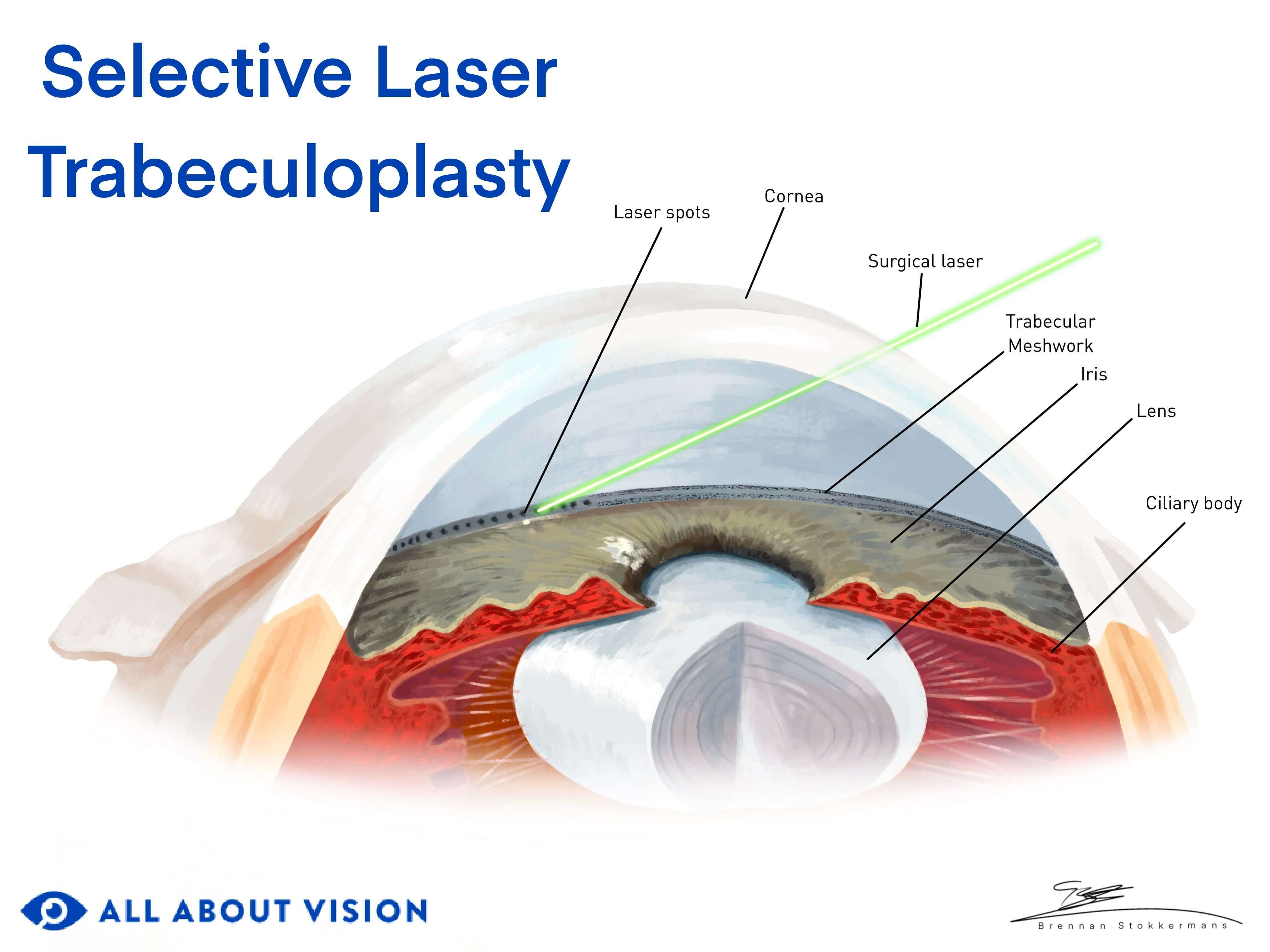
Laser Trabeculoplasty
Laser treatment helps improve fluid drainage from the eye and lower pressure. A glaucoma specialist may suggest it when medicines alone are not enough.
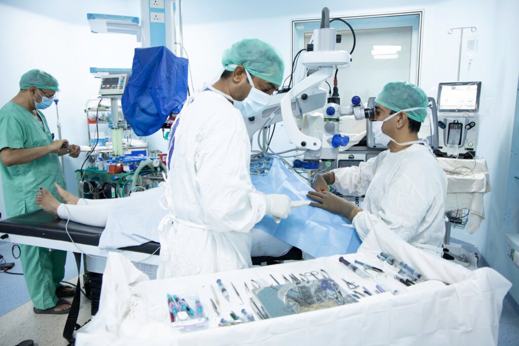
Glaucoma Filtering Surgery
When medicines or laser treatment cannot control eye pressure well, filtering surgery may create a new pathway for fluid to drain from the eye.
Combination Treatment
Some patients need more than one approach. Eye drops, laser or surgery may be combined depending on eye pressure, optic nerve health and disease progression.
Why Choose Viaan Eye and Retina Centre?
Should You Get Checked for Glaucoma?
Glaucoma can develop without obvious early symptoms. A check with a glaucoma specialist doctor can help assess your eye pressure, optic nerve and whether monitoring or eye glaucoma treatment is needed.
Protecting Your Vision Through Every Stage of Glaucoma
Glaucoma care is not a one time treatment. It starts with careful testing and continues with regular checks to protect your vision over time.
1. Detailed Eye Check
We begin by checking your eye pressure, optic nerve and overall eye health to understand whether glaucoma is present and how far it has progressed.
2. Understanding the Condition
Your glaucoma specialist doctor reviews the reports, explains what they mean and helps you understand the type and stage of glaucoma.
3. Choosing the Right Treatment
Your glaucoma specialist then plans the next step based on your eye pressure, optic nerve health, age and overall eye condition.
4. Starting Eye Glaucoma Treatment
Your eye glaucoma treatment may include eye drops, laser treatment, surgery or a combination of options depending on what your eyes need.
5. Regular Monitoring
Follow up visits help us keep a close watch on eye pressure and vision changes so treatment can be adjusted whenever necessary.

Your Vision Deserves Timely Glaucoma Care
Glaucoma can progress quietly. Speak with our glaucoma specialist doctor and take the right step before more vision is lost.