

Have you ever come across a situation where it becomes difficult for a trained and experienced medical professional to understand why a medical misdiagnosis has taken place? One of the major reasons can be such medical conditions that closely mirror one another and to such an extent that at times even the best eye specialists choose the wrong one.
The medical industry is dealing with different ways to cope up or reduce the chances of misdiagnosis. Also, from the patients’ perspective, it is important to ensure that their doctors are armed with professional expertise and requisite knowledge about their conditions and they can have full confidence in the decisions made by the doctors.
When we talk about the misdiagnosed retinal conditions, there are 3 common instances that usually take place:
- Macular Hole: The macula is the central area of the retina where light is sharply focused to produce the detailed colour vision and gives us the ability to see “20/20”. When a full-thickness defect develops in the macula, the condition is referred to as macular hole. A macular hole is a small gap at the centre which is common in people aged 60 and above. Initially, the person may feel blurriness and slight distortion in their straight-head vision but as it grows, a dark or blind spot may appear in your central vision.
Often the patients are told that they have a macular hole but what they actually have is the lamellar macular hole. The difference between the two is that a macular hole extends through all of the retina layers whereas a lamellar macular hole does not. A macular hole requires surgical treatment whereas a lamellar macular hole does not require surgical treatment unless the patient’s vision worsens.
- Retinoschisis: Retinoschisis is an eye disease caused by the abnormal splitting of the retina’s neurosensory layers. When it splits, tiny lumps called cysts form between the layers which damage the nerves and keep light signals from reaching the brain. Damaged nerves can make your vision blurry.
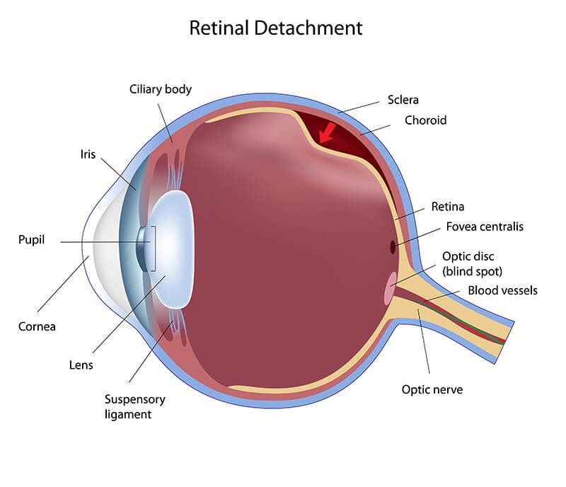
Sometimes, patients who were told that they have a retinal detachment actually have a condition called peripheral retinoschisis. The confusion occurs because both the conditions feature an elevated retina. A retinal detachment is sight-threatening and treated with surgery while a peripheral retinoschisis is not sight-threatening and is observed.
- Central Retinal Vein Occlusion: Central retinal vein occlusion (CRVO) is a condition in which the main vein that drains blood from the retina closes off partially or completely. The presence of scattered blot haemorrhages in all four quadrants can be seen in both central retinal vein occlusion (CRVO) and diabetic retinopathy which makes a medical misdiagnosis possible and unfortunately common. This can cause blurred vision and other problems with the eye.
The ultimate goal is to not let the patient lack confidence in their doctor or undergo any unnecessary treatment. This is why we, at Viaan Eye & Retina Centre, focus on delivering comprehensive eye care with cutting edge technology and personalized care. Our guiding philosophy is to provide ethical eye care with our latest technology & human intelligence.
Connect with us at +91-8448440121, if you have any queries. We at Viaan Eye & Retina Centre are happy to be of service.