


INVESTIGATIONS IN RETINAL DISEASES
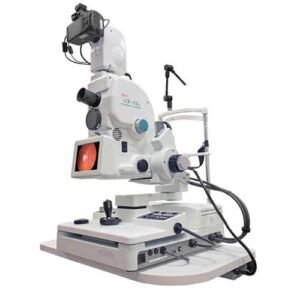
1. FUNDUS FLUORESCEIN ANGIOGRAPHY
A self-explanatory consent form, which explains the side effects in detail, is provided to the patients. Consent is required before the procedure. Patients can have a light meal before undergoing the procedure and must be accompanied by a family member.
Eye drops are administered to enlarge the pupils — this takes approximately 30-40 minutes. Patient is then be asked to sit still in front of the camera while a series of color photographs of eyes are taken. An injection (contrast) is then injected and more photographs are taken.
The test takes approximately 10-15 minutes.
It is a very safe procedure and serious side effects are uncommon. However, there is a rare possibility that a patient may have a reaction to the contrast. Some patients may experience slight nausea after the dye injection, but the feeling usually passes quickly.
Patients who are allergic to the dye can develop itching and a skin rash. These symptoms generally respond quickly to medications such as anti-allergics or steroids.
There is also a possibility of the dye going into the skin at the injection site, this may cause some discomfort or discoloring of the skin for several days. The fluorescein dye turns the patient’s urine orange and may slightly discolor the skin as well for a brief period.
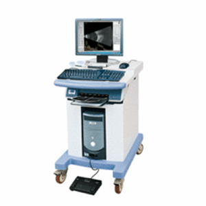
2. ULTRASONOGRAPHY OF POSTERIOR SEGMENT
A thick jelly is placed on the ultrasound probe, which is then gently placed on the eye over the lid. It is a safe and painless procedure, no X-ray exposure occurs in this test. Your doctor uses these echo pictures to arrive at a diagnosis, the probable future health of your eye, and a treatment plan. However, the test only reveals the structure of the inside of the eye.
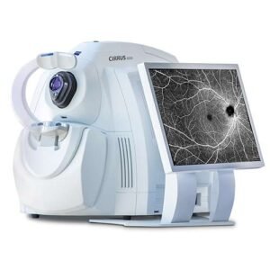
3. OPTICAL COHERENCE TOMOGRAPHY (OCT)
Optical coherence tomography is a non-invasive imaging test, which uses light waves to take cross-section pictures of patients’ retina.
With OCT, the ophthalmologist can see each of the retina’s distinctive layers. This allows mapping and measuring retinal thickness. These measurements help with diagnosis. OCT also guides treatment for retinal diseases, like age-related macular degeneration (AMD) , diabetic eye disease, macular edema.
The ophthalmologist may or may not put dilating eye drops . These drops widen the pupil and make it easier to examine the retina.
Patient sits in front of the OCT machine and rests head and chin on a support to keep them motionless. The equipment then scans the eye without touching it. Scanning takes about 5 to 10 minutes.
OCT is often used to evaluate disorders of the optic nerve as well. The OCT exam helps the ophthalmologist see changes to the fibers of the optic nerve. For example, it can detect changes caused by glaucoma.
OCT relies on light waves. It cannot be used with conditions that interfere with light passing through the eye. These conditions include dense cataracts or significant bleeding in the vitreous.
4. OPTICAL COHERENCE TOMOGRAPHY ANGIOGRAPHY (OCTA)
This test takes pictures of the blood vessels in and under the retina. OCTA is like fluorescein angiography, but is a much quicker test and does not use a dye (dyeless angiography). It can be safely done in patients allergic to fluorescein dye, and with renal disease (where dye/ contrast is contraindicated)
OCT and OCTA help diagnose many eye conditions, including:
macular hole
macular pucker
macular edema
age-related macular degeneration
central serous retinopathy
diabetic macular edema
abnormal blood vessels within/ underneath the retina
blood vessel blockage
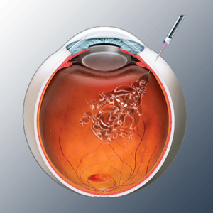
INTRAVITREAL INJECTIONS
a) Anti-VEGF treatment
Anti-VEGF (anti- Vascular Endothelial Growth Factor) injections are a group of medicines which reduce new blood vessel growth or oedema (swelling). They can be used to treat a number of eye conditions which cause new blood vessel growth or swelling under the macula area of the retina.
Currently anti-VEGF treatment is used for:
• ‘wet’ Age-related macular degeneration (AMD)
• Diabetic maculopathy
• Macular oedema caused by retinal vein occlusion.
• Myopic Choroidal Neovascularisation.
• Retinopathy of Prematurity. (ROP)
New blood vessel growth and macular oedema can also occur in other retinal conditions, so in the future anti-VEGF medications may be used in a wider range of situations.
Anti-VEGF treatments are given by an injection into the white portion of the eye and work by reducing the growth of new blood vessels and the oedema (swelling). Doing this can reduce the risk of scarring and damage to the retina caused by these new vessels, which in turn can help to avoid further sight loss and for some people cause an improvement in vision.
Depending on the condition of the patient, the ophthalmologist performs tests to decide whether treatment with antiVEGFs is required .
After examining the eye, the doctor may undertake further tests to have a clearer picture of the patient’s eye and symptoms.
Procedure
Anti-VEGF drugs are given as an injection into the vitreous (the jelly-like substance inside the eye). The injection is given through the white of the eye (the sclera). The injection needs to be given in a sterile way in an operating theatre.
The needle used for the injection is small and short and the injection itself only takes a few seconds. The patients have had an anaesthetic drop in their eye, before the procedure, so the injection feels like a small point of pressure on the eye.
After the injection, vision may be blurry for several hours because of the dilating eye drops; this should improve by the next day or the day after. The white of the eye may get red after the injection for a few days. Black swirls in the vision may be noticed for a few weeks (this is the drug floating in the vitreous gel). The eye usually feels comfortable by the next day. Some eye drops are given for a few days after the injection to prevent an infection.
The different AntiVEGFs available are
1. ACCENTRIX (RANIBIZUMAB)
2. EYLEA (AFLIBERCEPT)
3. PAGENAX (BROLUCIZUMAB)
4. AVASTIN (Bevacizumab, off label use)
5. RAZUMAB, RANIEYES, OCEVA ( RANIBIZUMAB BIOSIMILARS)
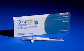
b) Steroids
Intravitreal steroids are used in some eyes with diabetic retinopathy, retinal vein occlusion and uveitis. Steroids help to reduce fluid leakage associated with these disorders.
The commonly used steroid is Dexamethasone Implant (Ozurdex), which is injected in the vitreous cavity just like any anti-VEGF drug.
c) Antibiotics
Antibiotic, anti fungal and antiviral drugs are injected in the vitreous cavity to treat patients with infections in the eye such as endophthalmitis and retinitis.